Images are for illustrative purposes only
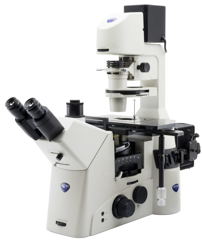
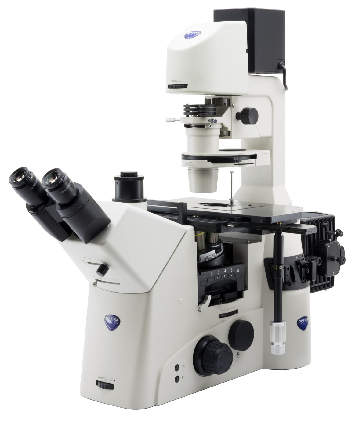
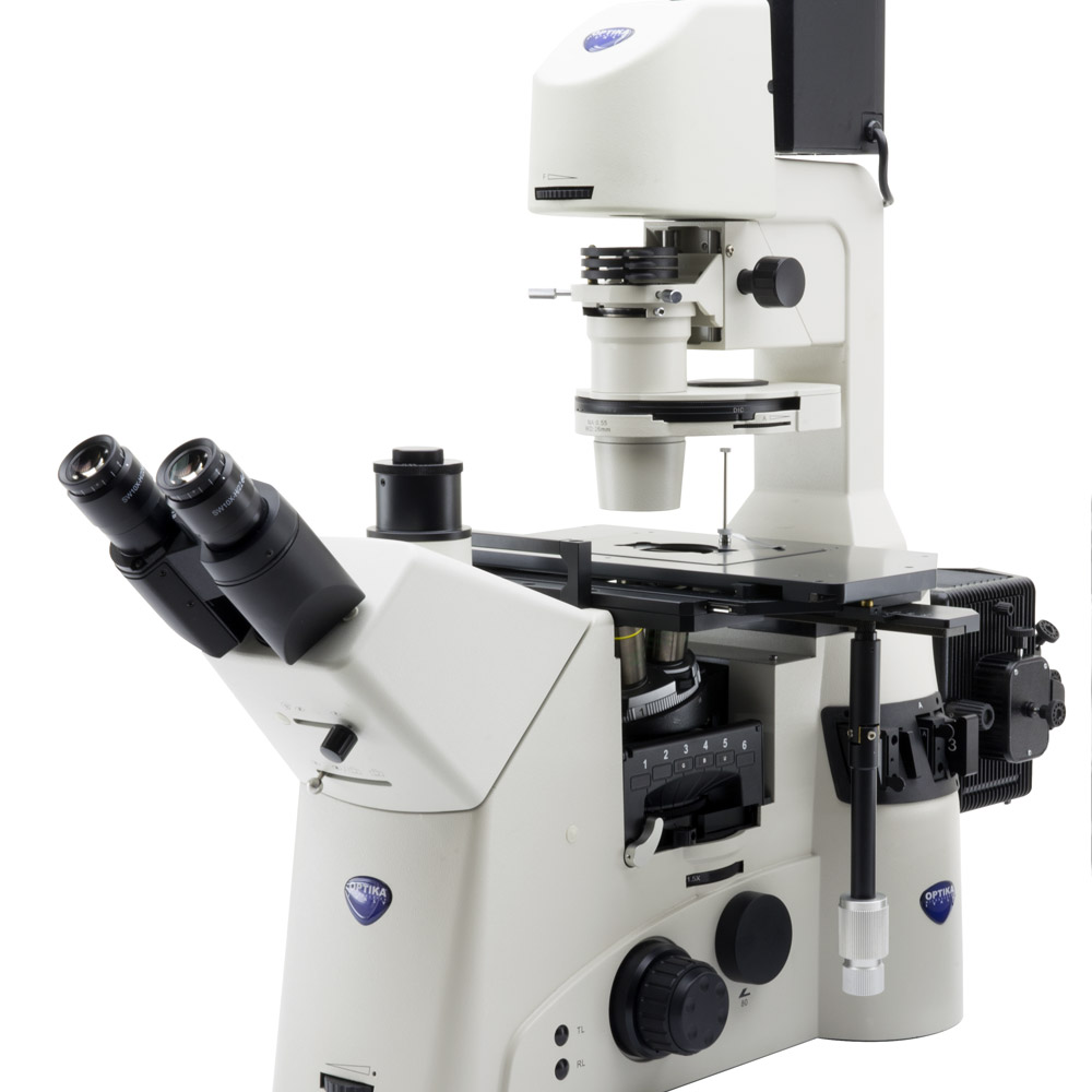
IM-7 represents the best of what Optika dedicates to the world of research.
This model was created to meet all the needs related to research in life science and designed to be
complemented by a series of packages dedicated to more advanced individual applications.
For all intents and purposes, IM-7 is to be considered as an inverted imaging platform, due to its
high expandability and state-of-the-art quality.
Top-level of optical equipment among our product range provides a sharp and clear view in any situation,
while top-level mechanical design offers sturdiness and long lifetime.
Images are for illustrative purposes only
BRIGHTFIELD
Transmitted brightfield illumination is one of the most commonly used observation
method in optical microscopy, and is ideal for fixed, stained specimens or other
types of samples having high natural absorption of visible light.
IM-7 is fitted with high-efficiency LED brightfield illuminator, for the best
outcome when using this technique.
Capsella middle embry – Brightfield


DIC
Differential Interference Contrast (DIC) is a microscopy technique that introduces
contrast to images of specimens which have little or no contrast when observed
using brightfield microscopy. The images produced using DIC have a pseudo
3D-effect, making the technique ideal for many applications.
DIC produces high resolution images with good contrast. It is best for
observing unstained samples.
Sphagnum pores g – DIC
FLUORESCENCE
The fluorescence microscopy is the most demanding
technique in biology and biomedical sciences, as well as in
materials science.
This method is capable to study organic and inorganic
samples thanks to primary fluorescence (auto-fluorescence)
or secondary (staining and labelling with fluorochromes)
IM-7 is tailored for applications in research, clinical and
pharmaceutical diagnostic field.
Fluorescence illuminators available as mercury lamp.
Cotton fibers – UV Fluorescence


PHASE CONTRAST
Phase-contrast microscopy is a particular technique applied in transparent, non-stainable, samples like culture of living cells, microorganisms, lithographic patterns, latex dispersions, fibers, asbestos and subcellular particles.
It reveals many cellular structures that are not visible with a simple brightfield microscope.
Diatoms – Phase contrast
Images are for illustrative purposes only

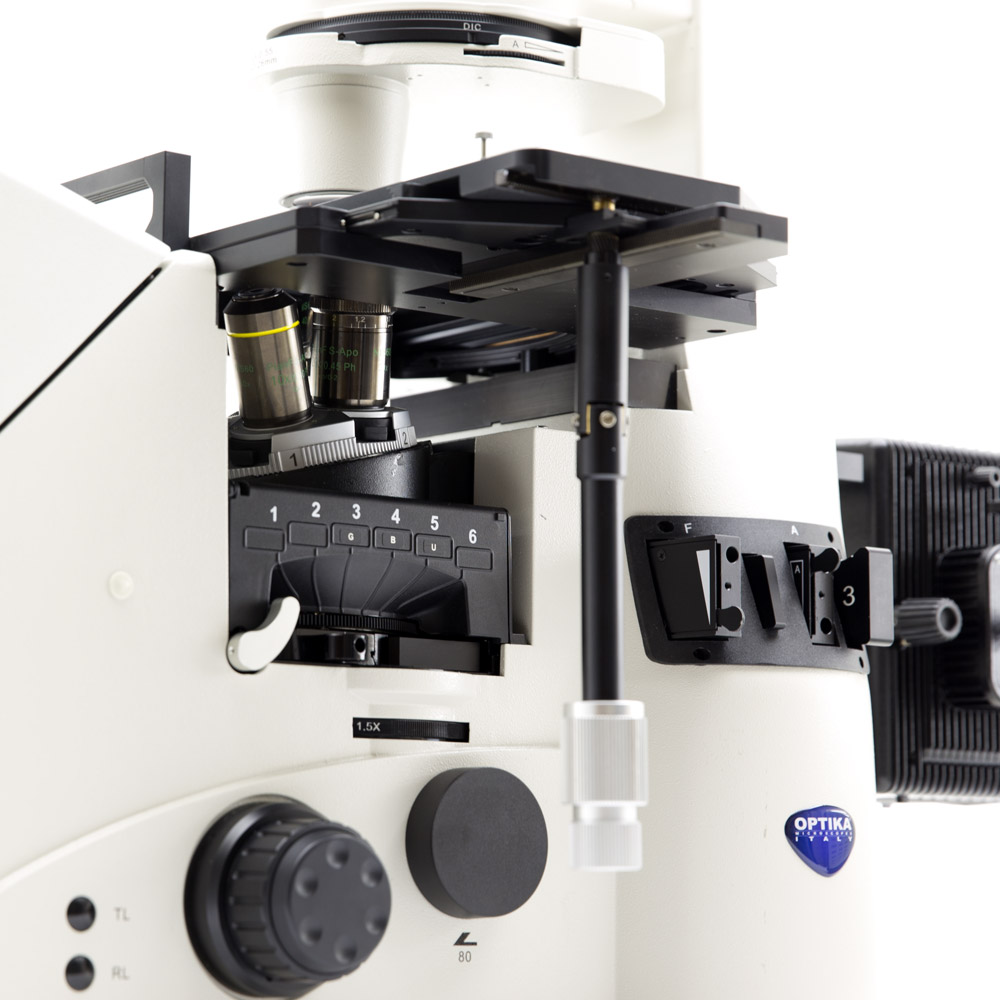
Revolving nosepiece for DIC and Fluorescence filter turret
The six-position nosepiece has a slot (in each of the six positions) for inserting DIC prisms.
The filter turret can hold up to six fluorescence filterblocks.
It is easily extractable, in order to facilitate the operation of inserting or replacing the filterblocks.
Mechanical stage and universal condenser
The wide 3-layer mechanical stage comes with several interchangeable plates for the use of Petri dishes, flasks and slides. The movement of the stage is controlled by a long tilting handle equipped with a pair of knobs for X/Y axes.
The universal condenser is a 6-position type, designed for brightfield, phase contrast and DIC.


Illuminator arm
The arm of the transmitted illuminator is backward tilting up to 30 degrees and it allows to use flasks and big bottles.
Main photo tube and Bertrand lens
The main photo tube located on the binocular head is easily controllable by using its control knob. 3 positions selectable:
100/0, 50/50, 0/100.
For phase contrast centering operations a Bertrand lens is available and it can be easily inserted by means of a dedicated knob.


Fluorescence equipment
A complete package of accessory dedicatd to Fluorescence technique is available as option.
Fluorescence filterblocks
In addition to the four standard fluorescence B-G-V-UV filterblocks, many others are available upon request to satisfy every kind of need.

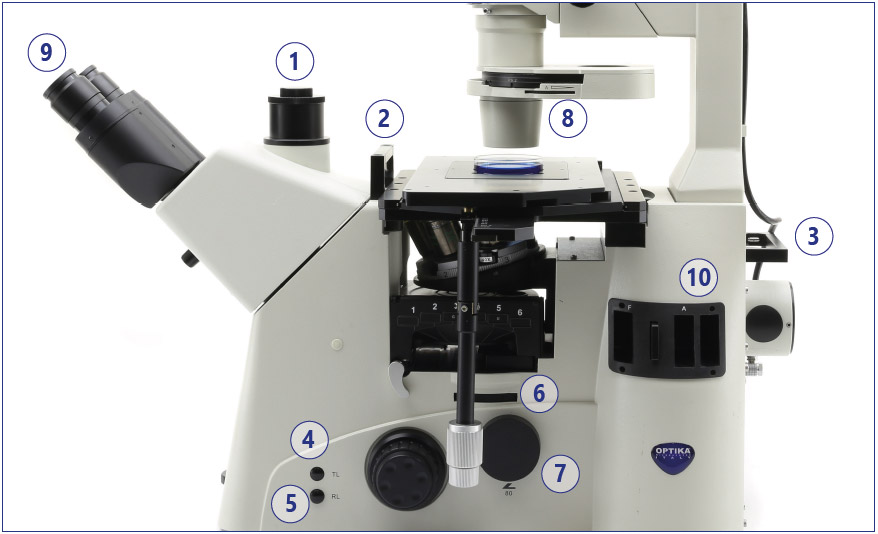
1 – Main photo tube, on binocular head
2 – Handle, for transportation
3 – Handle, for transportation
4 – Transmitted illuminator switch
5 – Fluorescence illuminator switch
6 – Magnification changer, 1x-1.5x
7 – Right side photo tube
8 – Universal condenser, for brightfield, PH and DIC
9 – Binocular head, with standard WF10x/22mm eyepieces or WF10x/25mm (optional)
10 – Slots for field diaphragm, aperture diaphragm, ND filters
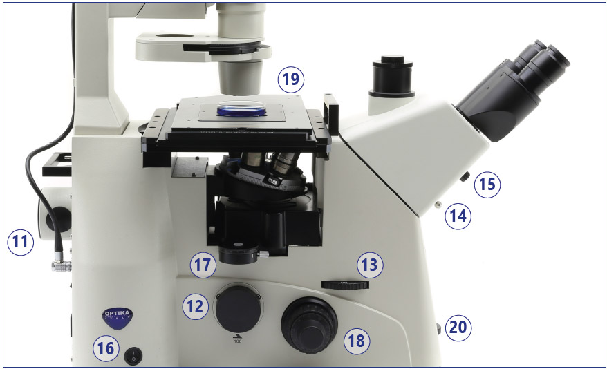
11 – Input port for fluorescence illuminator
12 – Left side photo tube
13 – Side photo tubes control (100/0 ; 20/80 ; 0/100)
14 – Main photo tube control (100/0 ; 50/50 ; 0/100)
15 – Bertrand lens insertion control
16 – Main switch
17 – Slot for analyzer
18 – Focusing knobs
19 – 3-layer mechanical stage
20 – Transmitted illuminator brightness control
IM-7 – Objectives/Application Packages
IM-7 is freely configurable in terms of objectives, by choosing among:
Infinity-corrected Semi-Apochromatic, Long Working Distance objectives, field flatness up to F.N. 25.
Objectives 20x, 40x, and 60x feature a correction collar in order to compensate for various thicknesses of cover glasses or different containers.
– M-1320 IOS U-PLAN F (SEMI-APO) PH 4x/0.13, W.D. 16.5 mm, Cover glass
– M-1321 IOS U-PLAN F (SEMI-APO) PH 10x/0.3, W.D. 7.4 mm, Cover glass 1.2 mm
– M-1322 IOS U-PLAN F (SEMI-APO) PH 20x/0.45, W.D. 7.5 – 8.8 mm, Cover glass 0 – 2 mm
– M-1323 IOS U-PLAN F (SEMI-APO) PH 40x/0.60, W.D. 3.0 – 4.4 mm, Cover glass 0 – 2 mm
– M-1324 IOS U-PLAN F (SEMI-APO) PH 60x/0.70, W.D. 1.8 – 2.6 mm, Cover glass 0.1 – 1.3 mm
– M-1358 IOS U PLAN APO Objective 60x/1.40 (Oil), W.D. 0.25 mm, Cover glass 0.17 mm
HBO Fluorescence package:
– M-1330 EPI Fluorescence internal attachment
– M-151.1 OSRAM 100W HBO high pressure mercury bulb
– M-1332 HBO Lamp house
– PS-HBO Optika 100W HBO power supply
– M-1334 6-position fluorescence filterbox turret
– M-1335 UV protector orange shield
– M-1336 B filterblock, filters included
– M-1337 G filterblock, filters included
– M-1338 V filterblock, filters included
– M-1339 UV filterblock, filters included
– M-1340 Aperture diaphragm slider
– M-1341 Field diaphragm slider
– M-1342 Slider with neutral density filter for HBO illumination
– M-1343 Empty fluorescence filterblock
M-1336: B (Blue)
Excitation filter (nm): 460 – 490
Dichroic cut-off mirror (nm): 505
Emission filter (nm): 515LP
M-1337: G (Green)
Excitation filter (nm): 510 – 550
Dichroic cut-off mirror (nm): 570
Emission filter (nm): 575LP
M-1338: V (Violet)
Excitation filter (nm): 385 – 425
Dichroic cut-off mirror (nm): 440
Emission filter (nm): 455LP
M-1339: UV (Ultraviolet)
Excitation filter (nm): 325 – 375
Dichroic cut-off mirror (nm): 415
Emission filter (nm): 435LP
DIC – Differential Interference Contrast package:
– M-1350 DIC Prism for 10x
– M-1351 DIC Prism for 20x
– M-1352 DIC Prism for 40x
– M-1353 DIC prism for 60x
– M-1354 DIC 1 prism 10x for condenser
– M-1355 DIC 2 prism 20x-40x for condenser
– M-1356 Slider with rotating analyzer
PH – Phase Contrast package
– M-1348 4X Phase ring
– M-1349 60X Phase ring