Striking Colors & 3D Shadowed-Like Appearance
DIFFERENTIAL INTERFERENCE CONTRAST (DIC)
Differential Interference Contrast (DIC) microscopy is a contrast technique that allows transparent structures to be visualized by exploiting changes in refractive index. Unstained biological specimens usually do not alter the amplitude of the incident light, i.e., they are phase specimens.
Usually, detectors (eye, camera etc.) are sensitive only to the intensity. Hence, visualization of phase specimen or reliefs requires some special method to convert phase information to intensity information, with the high contrast and 3-D relief effect that is characteristic of DIC.
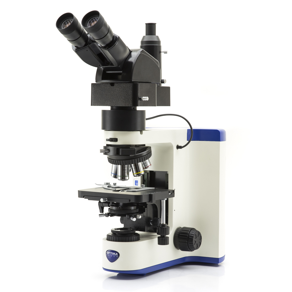
Köhler DIC (Differential Interference Contrast) on Transmitted Light – M-1198
DIC microscopy is ideal for unstained living specimens (like cultured cells, embryos, blood smears, diatoms, protozoa) and provides morphological information without the need to use potentially toxic dyes and fluorophores.
DIC is, however, often used in association with fluorescence to reveal morphological features of the specimen.
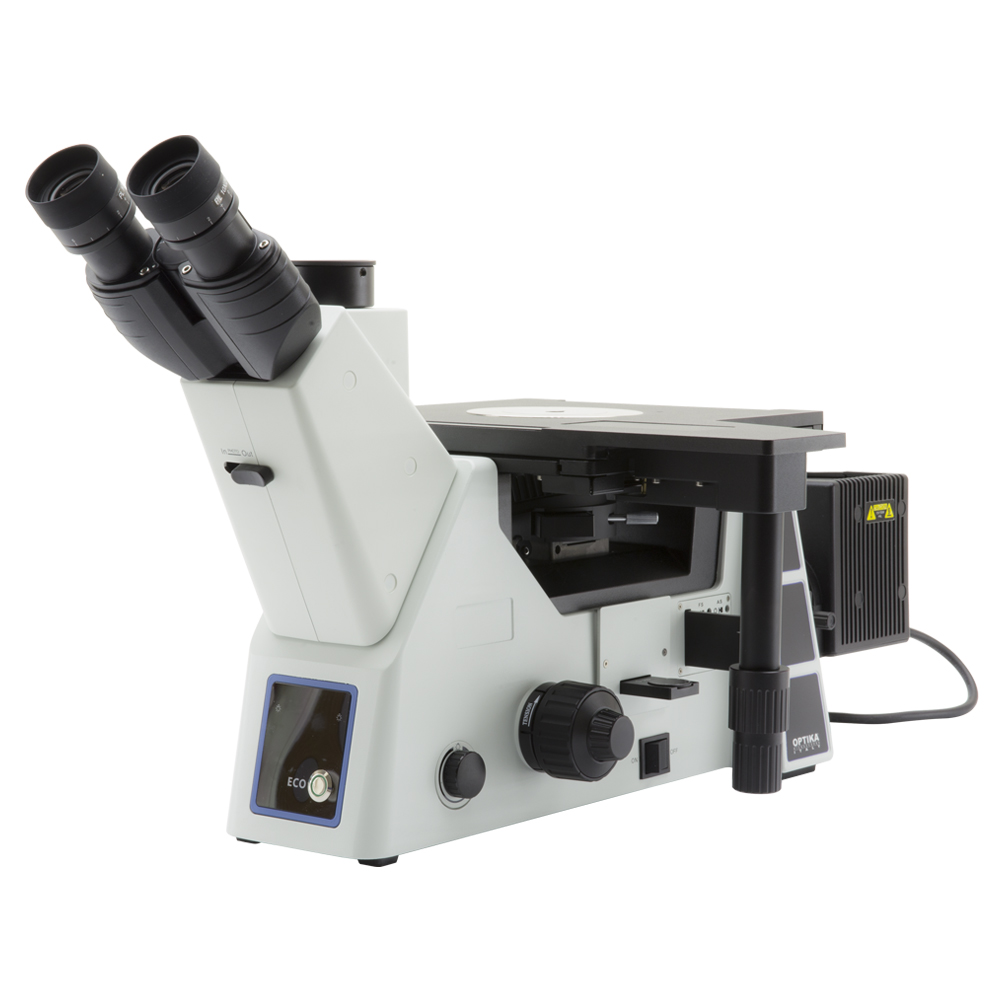
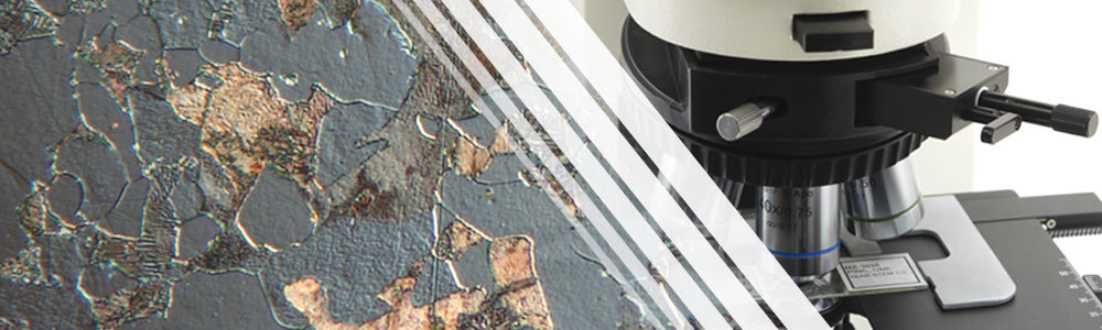
Nomarski DIC (Differential Interference Contrast) on Reflected Light – M-870
In metallography applications, images created in reflected light DIC can be interpreted as a true three-dimensional representation of the surface geometry: a clear distinction can be realized between raised and lowered regions in the specimen. It is also used for the examination of polymers and other materials.