OPTIKA News
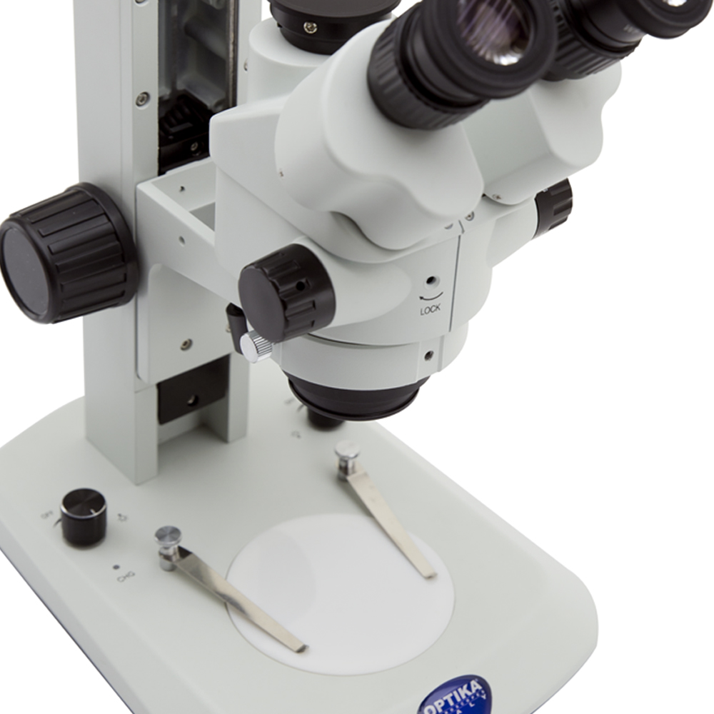
SZ Series
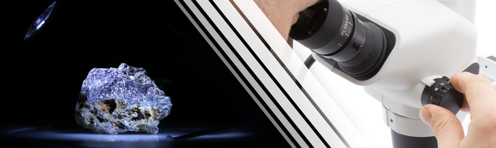
New Line of Stereomicroscopes
AN INCREDIBLE VARIETY OF OVER 90 CONFIGURATIONS
OPTIKA has decided to drastically enlarge the variety of stereozoom microscopes with the introduction of the new SZ Series, with more than 90 possible configurations!
Just three simple steps are needed:
1. Identify The Most Suitable Head For Your Purposes
SZX Heads – Excellent Price/Performance Ratio For Any Laboratory
» Ideal for universities, experts & common routine lab requirements
» 22 mm field number and large working distance (up 110 mm)
» 6.72:1 zoom ratio – zoom magnification from 6.7x to 45x
» Cost-effective solution for diversified applications
SZX-A Heads – Advanced Analysis With High Power Magnification
» Purposely designed for particularly performing zoom conditions
» 23 mm field number and large working distance (up 110 mm)
» 8.46:1 zoom ratio – zoom magnification from 6.5x to 55x
» High magnification change from overview to tiny details
SZO Heads – Addressed For Extreme Reliability & Repeatability
» Ensuring the sharpest vision, high productivity, repetitive analysis
» 23 mm field number and large working distance (up 110 mm)
» 6.72:1 zoom ratio – zoom magnification from 6.7x to 45x
» Multi-position click-stop – no need to move eyes from eyepieces

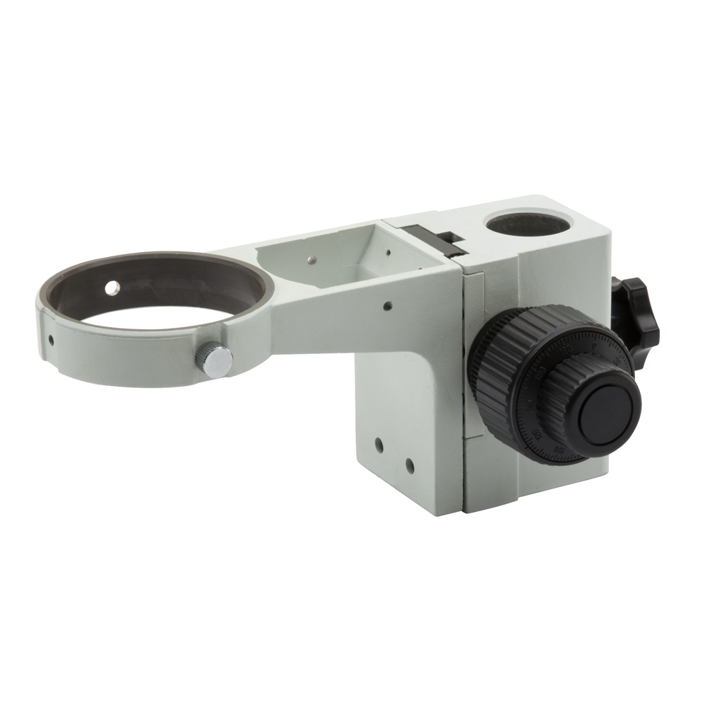
2. Select The Ideal Focusing System
Different Challenges Requires Different Focusing System
» Entry-level coarse focusing to raise or lower the head to focus
» Coaxial coarse and fine focusing results in easy, precise conditions
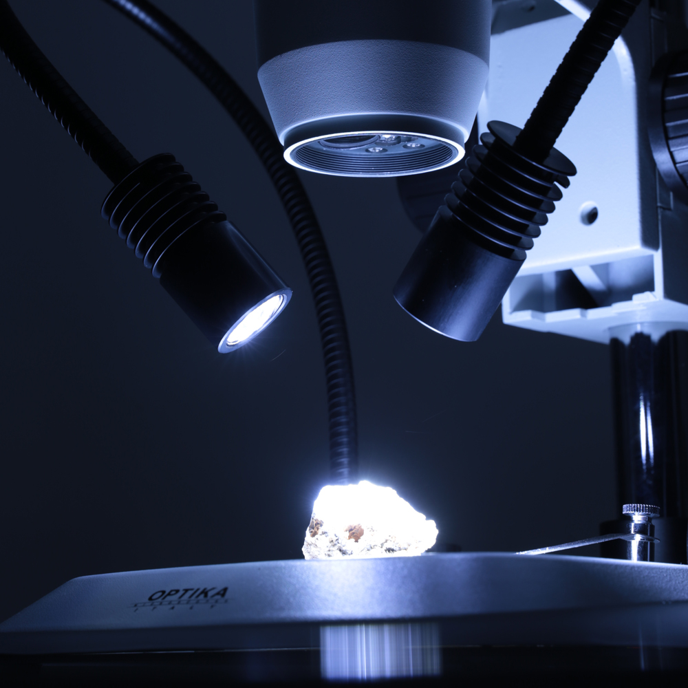
3. Combine The Best Fit Stand
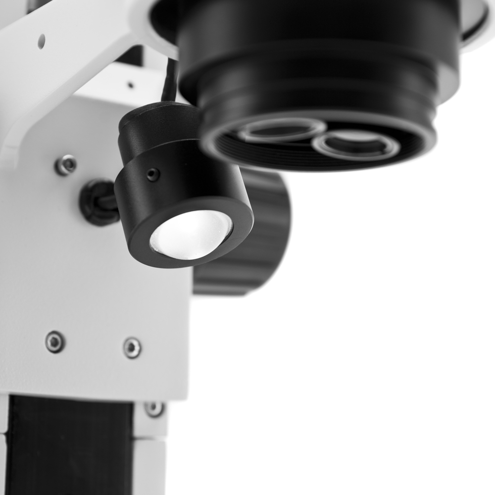
Plenty Of Smart & Innovative Light-Related Technologies – X-LED
» Large, ultra-flat stands with excellent stability (w/ or w/o light)
» Universal, 360° rotating, tilting overhanging stands for large sample
» State-of-the-art illumination system for incomparable light intensity
» Top-class X-LEDT transmitted light, with geometrically arranged LEDs
» Freely orientable, flexible double gooseneck X-LED3 incident light
Contact us immediately to get more information about SZ Series !
OPTIKA Academy
Sales & Applications Training Course
REGISTRATIONS ARE NOW OPEN!
OPTIKA Microscopes offers tons of great sales arguments that will be a perfect fit for your customers…but are you sure to know all of them (including the more “secrete” ones)?
Come and visit us in our headquarters in Bergamo, Italy to take a look at the different operations of the company. At the same time, you will increase your confidence in our products and receive informative tips helping you to convince more and more customers on the spot to join OPTIKA users’ community. These days will be a great personal development, sharing and networking opportunities with a focus on improving and updating your microscopy knowledge.
On top of that, good Italian food and a friendly staff is looking forward to welcoming you in this pleasant experience.
Registrations are now open for:
Sales & Application Training Course – November 25-27, 2019
Language: english
Send your application to your usual OPTIKA contact now and impressively enhance the chance of your success!
Spaces are limited: book now to prevent disappointment and wait for OPTIKA confirmation.
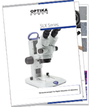
SLX Series
New Line of Cordless Stereomicroscopes
PROFESSIONAL FEATURES AVAILABLE TO… WELL, EVERYONE!
Discover now the brand new OPTIKA SLX Series, valuable configurations of cordless and modern 3D Greenough stereomicroscopes with several key features and ideal for a variety of applications, including dissection, biology, entomology, anatomy, chemistry and material science among the others.
High eyepoint eyepieces, 21mm FN
Designed in such a way that the exit pupil is further away from the eye lens than standard eyepieces, being are well suited for eyeglasses wearers
6.43:1 zoom ratio – zoom magnification from 7x to 45x
Purposely designed for professional routine inspections, the total magnification can be even extended to 135x with 20x eyepieces and 1.5x additional lens, obtaining an excellent results in this class
Ultra-flat base with Ø 100 mm disc for diffused transmitted light
A new level of ergonomy and comfort is achieved during operations, with the ultra-flat base of only 3 cm height to ensure smooth specimen movement and the Ø 100 mm for top class diffusion of the transmitted light
The longest autonomy on the market ensured by EcoLED™
OPTIKA has re-designed illumination in microscopy, once again: a special coating process on optics combined with a new, higher ratio between low consumptions and ultra-efficiency has addressed us to top brightness levels, providing over 20 years of use
Cordless use, totally independent from mains/batteries connection
All models work with or without the batteries in place and are provided with three NiMH rechargeable batteries for the longest autonomy in outdoor use (12-hour autonomy, at medium intensity)
Contact us immediately to get more information about SLX Series!
B-60 Series
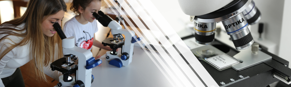
New Line of Cordless Educational Microscopes
IDEAL TO START EXPLORING!
Perfect for the first experiences, OPTIKA B-60 Series represents the most modern cordless microscope ideal for students.
Slim and easy to carry, every detail has been carefully thought out to let the user work with enhanced comfort and intuitiveness compared to any other solutions in this class.
Available in several configurations to meet the most diversified needs, B-60 is powered by a bright LED to ensure clear observations over 20 years of use and totally independent from the mains thanks to the rechargeable batteries.

The StagErase™ is something you’ve never seen before: the new, revolutionary stage of OPTIKA B-60 is coated with a special painting to reduce accidental scratches caused by the slides to the minimum and facilitate their removal.
The ergonomic design includes an arm / wrist rest support to let students be relaxed and stay relaxed when using the microscope, reducing the fatigue during use and increasing the performance as a result.
ARABLAB 2019
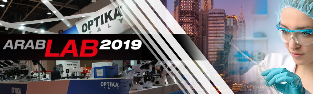
SAVE THE DATE – March, 12-14
ARABLAB 2019, UAE – Booth 954, Hall S1
Do not miss the chance to visit OPTIKA Microscopes booth at ARABLAB 2019 to learn about our latest exciting developments, including the new B-510 & IM-5!
Where: Booth 954, Hall S1, ARABLAB, Dubai (UAE)
When: March, 12-14.
Save your time: write now to your usual contact in OPTIKA and schedule an appointment.
Looking forward to meeting you.
10-YEAR WARRANTY
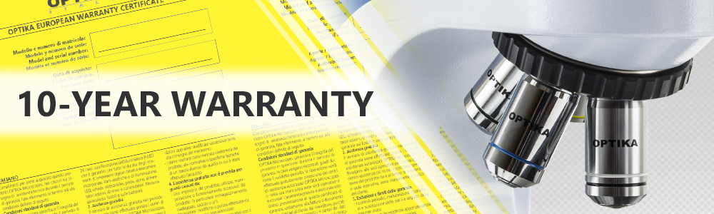
The Longest Coverage in the Industry
IT IS TIME TO TAKE A DECISION YOU WILL THANK YOURSELF FOR LATER!
From now, OPTIKA is offering to customers a 10-year warranty on optical and mechanical parts.
The warranty period begins on the date of invoice and applies worldwide.
The extended warranty applies starting from today!
Warranty for digital components of microscopes, electronic/eletrical parts and illumination is 2-year.
X-LEDs is warranted for the entire life of the instruments.
For further info, please refer to the “General Sales Conditions” listed in the OPTIKA price list.
The 10-year warranty is voided if changes/modifications have been done by customers and applies for instruments which have been used as intended.
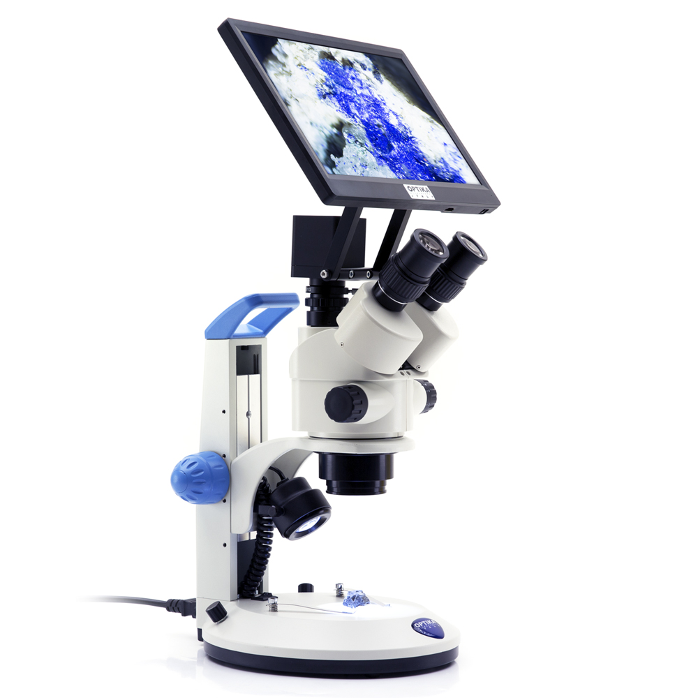
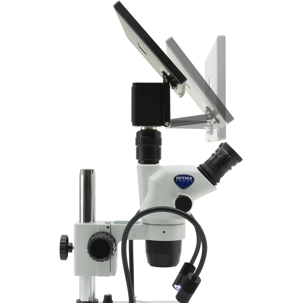
C-HESC & C-HPSC
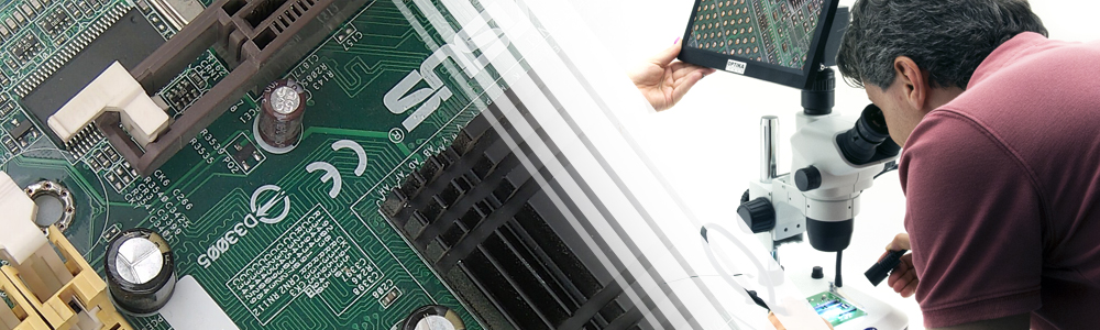
New Line of Digital Products
Compatible with any biological or stereo microscope, these HDMI cameras are equipped with the latest technology ensuring great contrast and excellent color reproduction for beautiful true-to-life.
The specimen is displayed on the monitor which can be tilted and positioned according to user preferences, sitting in a comfortable and relaxed position (especially after extensive use) for enhanced ergonomy.
Productivity is drastically improved since samples will be processed much faster than with a conventional system.
On top of that, more people can enjoy the image output simultaneously for discussion groups.
Still undecided? Do not hesitate to contact us!
DIC Microscopy
Striking Colors & 3D Shadowed-Like Appearance
DIFFERENTIAL INTERFERENCE CONTRAST (DIC)
Differential Interference Contrast (DIC) microscopy is a contrast technique that allows transparent structures to be visualized by exploiting changes in refractive index. Unstained biological specimens usually do not alter the amplitude of the incident light, i.e., they are phase specimens.
Usually, detectors (eye, camera etc.) are sensitive only to the intensity. Hence, visualization of phase specimen or reliefs requires some special method to convert phase information to intensity information, with the high contrast and 3-D relief effect that is characteristic of DIC.
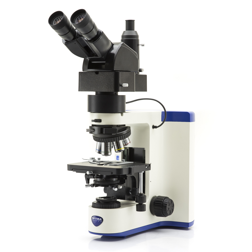
Köhler DIC (Differential Interference Contrast) on Transmitted Light – M-1198
DIC microscopy is ideal for unstained living specimens (like cultured cells, embryos, blood smears, diatoms, protozoa) and provides morphological information without the need to use potentially toxic dyes and fluorophores.
DIC is, however, often used in association with fluorescence to reveal morphological features of the specimen.
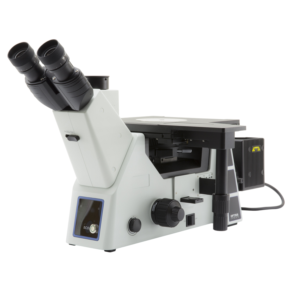
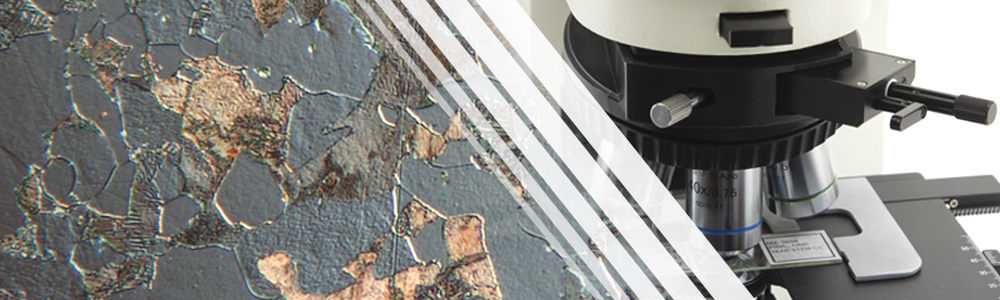
Nomarski DIC (Differential Interference Contrast) on Reflected Light – M-870
In metallography applications, images created in reflected light DIC can be interpreted as a true three-dimensional representation of the surface geometry: a clear distinction can be realized between raised and lowered regions in the specimen. It is also used for the examination of polymers and other materials.
B-810
New Upright Microscopes
B-810 UPRIGHT MODULAR MICROSCOPES
OPTIKA B-810 helps you work in a comfortable way during extended periods of use. Versatile, robust, durable and sturdy, B-810 offers premium quality optics, the state-of-the-art, exclusive X-LED3 illumination system, designed by OPTIKA, for true colors, and the Köhler diaphragm. Ready for digital imaging, B-810 will be the perfect assistant for your routine applications requiring brightfield and phase contrast techniques.
Transmitted brightfield illumination is one of the most commonly used observation method in optical microscopy, and is ideal for fixed, stained specimens or other types of samples having high natural absorption of visible light.
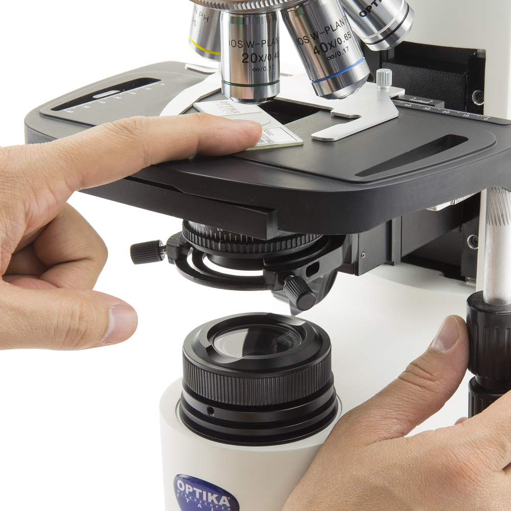
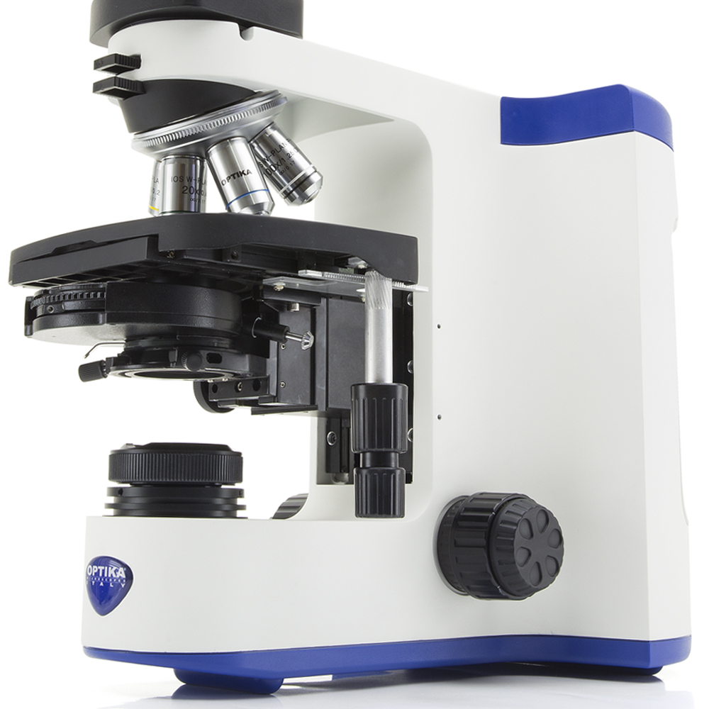
Phase-contrast microscopy is a particular technique applied in transparent, non-stainable, samples like culture of living cells, microorganisms, lithographic patterns, latex dispersions, fibers, and subcellular particles, revealing many cellular structures that are not visible by using brightfield.
ACHEMA 2018
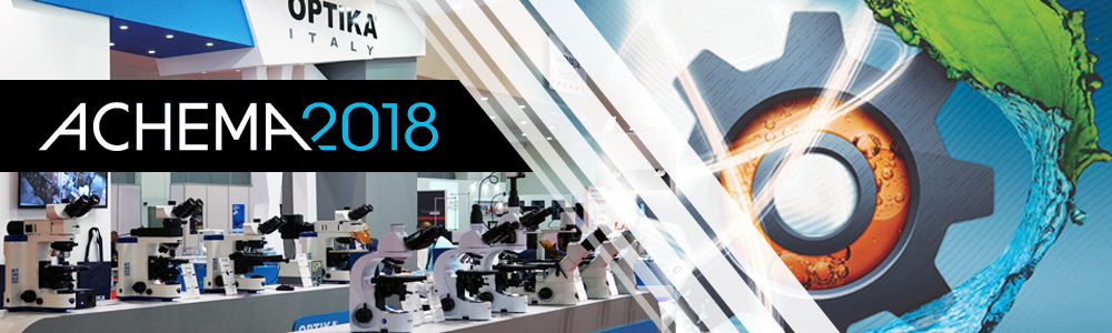
SAVE THE DATE – June, 11-15
ACHEMA 2018, Germany – Booth H61, Hall 4.2
Do not miss the chance to visit OPTIKA Microscopes booth at ACHEMA 2018 to learn about our latest exciting developments, especially the brand new B-510 & IM-5!
Where: Booth H61, Hall 4.2, ACHEMA, Frankfurt am Main (Germany)
When: June, 11-15.
Save your time: write now to your usual contact in OPTIKA and schedule an appointment.
Looking forward to meeting you.